Anatomy Of The Upper Chest Area ~ Muscles Of The Trunk Anatomy Diagram Pictures Kenhub. Anatomy of the upper chest area the subclavian artery supplies portions of the chest cavity and chest wall and portions of the shoulder girdle. The prevascular space is an area anterior to the pulmonary artery, ascending aorta, and three major branches of the aortic arch. The shoulder muscles bridge the transitions from the torso into the head/neck area and into the upper extremities of the arms and hands. The abdomen (commonly called the belly) is the body space between the thorax (chest) and pelvis. The sternum, or breastbone, is a flat bone at the front center of the chest.
Jocelyn and briac are studying for an anatomy test. The shoulder joint is the junction between the chest and the upper extremity. Chest from left to right is the. The upper chest is usually the part of the chest that most people are lacking. The diaphragm forms the upper surface of the abdomen.
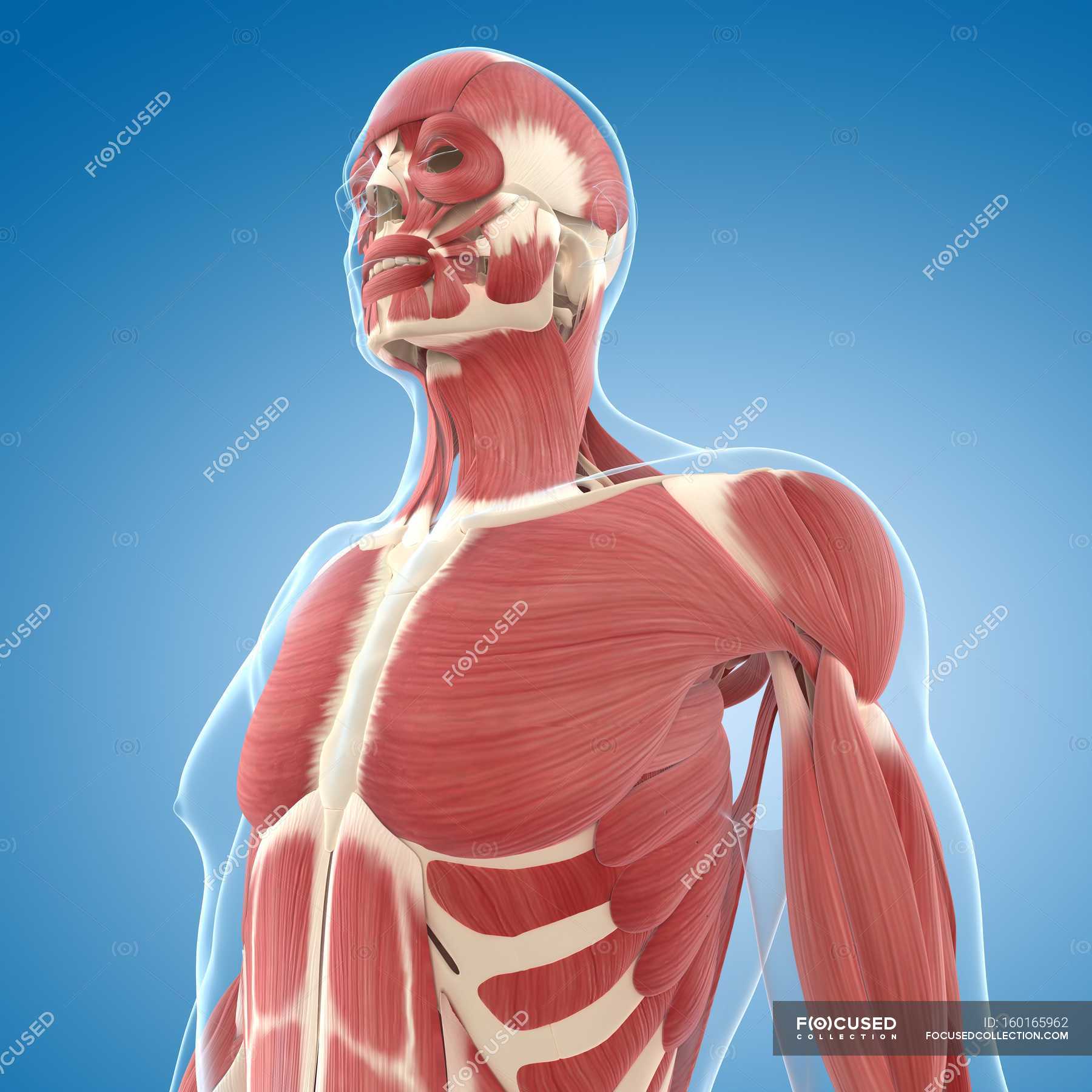
Upper Chest Musculature Male Musculature Anatomy Stock Photo 160165962 from st.focusedcollection.com Its meaning is simple, but it is a very complicated area to describe in words. Related posts of anatomy of the chest area. Your sternum protects the organs of your torso from injury and also serves as a. The rest of the points in this meridian are on the arm. It's also sometimes referred to as the breastbone. Not much to see here. Jocelyn says the upper thigh is proximal to the hip when compared to the knee. The larynx is made up of a total of nine different types of cartilages.
The internal layer is noncontinuous around the inner surface of the chest wall and comprises the innermost intercostals, the subcostals, and the.
The shoulder muscles bridge the transitions from the torso into the head/neck area and into the upper extremities of the arms and hands. The upper part of the larynx made up of tissues is known as the supraglottis while subglottis refers to the tissues at the bottom that connect the trachea and the larynx. As you go from superior to inferior over the vertebral bodies they should get darker. It is where the vocal cords are located. Jocelyn and briac are studying for an anatomy test. The upper parts of the chest are also found in various parts of the body. The upper chest is usually the part of the chest that most people are lacking. For that reason, and because of the dexterity of the shoulder joint itself, the musculature of the shoulder is. Chest wall (anterior view) therefore, the thorax can be defined as consisting of the thoracic cavity, its contents including the primary organs of the respiratory and cardiovascular systems, and the wall that surrounds it. Anatomy of the upper chest area synopsisthe chest wall like other regional anatomy is a wondrous fusion of form and function. The sternum, or breastbone, is a flat bone at the front center of the chest. Thoracic cavity, also called chest cavity, the second largest hollow space of the body.it is enclosed by the ribs, the vertebral column, and the sternum, or breastbone, and is separated from the abdominal cavity (the body's largest hollow space) by a muscular and membranous partition, the diaphragm.it contains the lungs, the middle and lower airways—the tracheobronchial tree—the heart. Anatomy of right side chest pain.
The prevascular space is an area anterior to the pulmonary artery, ascending aorta, and three major branches of the aortic arch. Your sternum protects the organs of your torso from injury and also serves as a. Chest from left to right is the. Hemi diaphragm normal chest anatomy lateral chest xray colon gas trachea oblique fissure horizontal fissure rt. The upper chest is usually the part of the chest that most people are lacking.
What Are The Most Common Causes Of Pectoral Muscle Pain from images.infobloom.com The ribs and sternum make up what is called the 'ribcage.' the ribcage protects the lungs, blood vessels, and heart,. The pectoral region is located on the anterior chest wall. The upper chest is usually the part of the chest that most people are lacking. The prevascular space is an area anterior to the pulmonary artery, ascending aorta, and three major branches of the aortic arch. Area surrounding the heart, where the lungs are. The upper parts of the chest are also found in various parts of the body. Upper thorax meaning is used for identifying or discussing the chest area. Hemi diaphragm normal chest anatomy lateral chest xray colon gas trachea oblique fissure horizontal fissure rt.
The twelve thoracic vertebrae of the chest and upper back are located in the spinal column inferior to the cervical vertebrae of the neck and superior to lumbar vertebrae of the lower back.
Related posts of anatomy of the chest area. The diaphragm forms the upper surface of the abdomen. Which terms describe the area that she could be referring to? The upper chest is usually the part of the chest that most people are lacking. It is the final set of ribs that makes up the back and the top part of the rib cage. The pectoral region is located on the anterior chest wall. Chest from left to right is the. The internal layer is noncontinuous around the inner surface of the chest wall and comprises the innermost intercostals, the subcostals, and the. See chest anatomy stock video clips. A chest ultrasound is a noninvasive diagnostic exam that produces images, which used to assess the organs and structures within the chest, such as the lungs, mediastinum (area in the chest containing the heart, aorta, trachea, esophagus, thymus, and lymph nodes), and pleural space (space between the lungs and the interior wall of the chest). No need to register, buy now! Abbie went to the hospital complaining of chest pain. Chest wall (anterior view) therefore, the thorax can be defined as consisting of the thoracic cavity, its contents including the primary organs of the respiratory and cardiovascular systems, and the wall that surrounds it.
In humans and other hominids, the thorax is the chest region of the body between the neck and the abdomen, along with its internal organs and other contents. Nerves of the chest and upper back. It is mostly protected and supported by the rib cage, spine, and shoulder girdle. Its meaning is simple, but it is a very complicated area to describe in words. The larynx is made up of a total of nine different types of cartilages.
Chest Pain Is It Heart Attack Or Nutcracker Esophagus Health Essentials From Cleveland Clinic from health.clevelandclinic.org The twelve thoracic vertebrae of the chest and upper back are located in the spinal column inferior to the cervical vertebrae of the neck and superior to lumbar vertebrae of the lower back. Briac says the upper thigh is inferior to the hip. In humans and other hominids, the thorax is the chest region of the body between the neck and the abdomen, along with its internal organs and other contents. Anatomy of the chest and shoulder, anatomy of the chest organs, anatomy of the chest wall, anatomy of the chest wall and pleura, anatomy of upper chest area, human. Chest wall (anterior view) therefore, the thorax can be defined as consisting of the thoracic cavity, its contents including the primary organs of the respiratory and cardiovascular systems, and the wall that surrounds it. It's also sometimes referred to as the breastbone. A second joint in the shoulder is the junction of the collar bone with the shoulder blade, called the. Find the perfect chest anatomy stock photo.
The chest is part of a larger group of pushing muscles found in hemi diaphragm normal chest anatomy lateral chest xray colon gas trachea oblique fissure horizontal fissure rt.
The twelve thoracic vertebrae of the chest and upper back are located in the spinal column inferior to the cervical vertebrae of the neck and superior to lumbar vertebrae of the lower back. At the level of the pelvic bones, the abdomen. The abdomen (commonly called the belly) is the body space between the thorax (chest) and pelvis. The larynx is made up of a total of nine different types of cartilages. Not much to see here. Nerves of the chest and upper back. The ribs and sternum make up what is called the 'ribcage.' the ribcage protects the lungs, blood vessels, and heart,. It's also sometimes referred to as the breastbone. Anatomy of right side chest pain. Each one spans half of the upper chest, and has attachment points on the sternum (breastbone), ribs, clavicle (collarbone), and humerus (long bone of your upper arm). The nervous system of the thorax is a vital part of the nervous system as a whole, as it includes the spinal cord, peripheral nerves, and autonomic ganglia that communicate with and control many vital organs. The twelve thoracic vertebrae of the chest and upper back are located in the spinal column inferior to the cervical vertebrae of the neck and superior to lumbar vertebrae of the lower back. The anatomy of the sternum.
Share :
Post a Comment
for "Anatomy Of The Upper Chest Area ~ Muscles Of The Trunk Anatomy Diagram Pictures Kenhub"


Post a Comment for "Anatomy Of The Upper Chest Area ~ Muscles Of The Trunk Anatomy Diagram Pictures Kenhub"